Breast Lumps Symptoms, Risk Factors & Treatment | Diseases List A-Z
Breast lumps are abnormal tissues that grow in the breast.
The tissue is not cancerous and does not pose a risk of triggering health problems in the sufferer.
This condition is more at risk for women than men.
Breast Lump Symptoms The symptoms experienced by people with lumps in the breast include: The lump is less or more than 5 centimeters in size.
This lump can grow larger over time.
The lumps enlarge before the menstrual cycle and return to their original size after menstruation is over.
The lump may feel soft, rubbery, or solid.
Lumps appear singly or in groups, on one side or both breasts at once.
The lump is round or oval in shape, can be moved or fixed.
Swelling of the breast.
Breasts feel tight.
Breasts feel warm to the touch.
The difference in size between the two breasts.
The nipples discharge clear or cloudy fluid.
Nipples feel itchy or sensitive.
Meanwhile, symptoms of lumps that need to be watched out for include: The lump is getting bigger.
The lump feels solid and does not move when moved.
The lump does not disappear after the menstrual cycle occurs.
The time ranges from more than 4 to or 6 weeks.
Breast skin becomes red, hard, or wrinkled like orange peel.
Breast bruising for no apparent reason.
Nipples turn inward or have an abnormal shape.
Nipples are bleeding.
A new lump appears.
A lump appeared in the armpit.
Causes of Breast Lumps The causes of lumps in the breast include: 1.
Fibroadenoma Fibroadenoma is a benign tumor that is prone to occur in women aged 20 to 30 years.
The tumor is formed from changes in breast tissue and connective tissue.
The trigger is changes in the hormone estrogen or the use of birth control pills before the age of 20.
2.
Fibrocystic breast Fibrocystic breasts are abnormal fibrous tissue growth in the breasts, making them more prominent than fatty tissue.
This disorder is common in women aged 30 to 50 years.
The trigger is changes in the hormone estrogen in the menstrual cycle.
Cysts are fluid-filled lumps that are round or oval in shape, soft in texture, and sometimes feel solid when touched.
The trigger can be a buildup of fluid in the mammary glands due to hormonal changes in the menstrual cycle.
Lipoma is a fatty lump that grows slowly under the skin of the breast.
This condition is prone to be experienced by women aged 40 to 60 years.
The risk is higher if there is a history of lipoma in the family.
5.
Mastitis Mastitis is an inflammation of the breast tissue experienced by breastfeeding mothers.
This disorder causes an abscess (collection of pus) in the breast tissue.
Bacterial infection of the breast tissue and blockage in the ducts (the channels that carry breast milk from the mammary glands to the nipple) can trigger this condition.
6.
Fat necrosis This is damage to the fatty glands in the breast, which generally occurs due to injury, after surgery, or after receiving radiation therapy to the breast.
7.
Intraductal papilloma Intraductal papilloma is a benign tumor that forms in the ducts.
These are the channels that carry milk from the mammary glands (lobule) to the nipple.
The tumor is made up of fibrous tissue, glands, and blood vessels.
The disease generally affects women aged 35 to 55 years.
Breast Lump Trigger Factors Factors that increase the risk of breast lumps include: Genetic history inherited from parents.
Hormonal changes in the body.
Using birth control pills.
Wounds resulting from injuries resulting from blunt force trauma.
Eating unhealthy foods, such as red meat and processed or fast foods.
Excessive consumption of alcoholic beverages.
Have a smoking habit.
Breast Lump Diagnosis First of all, the diagnostic step is done with a complete medical interview followed by a physical examination of the breast.
Then, the doctor will perform supporting tests to confirm the disease, such as: 1.
Mammography Mammography is an X-ray of the breast.
During the examination, the breast is pressed to make the breast tissue more visible.
This procedure can detect abnormalities, such as tumors, calcium deposits, or dense tissue in the breast.
2.
Ultrasonography (USG) Ultrasound is an examination that uses sound waves to display images on a monitor screen.
This procedure aims to distinguish solid lumps from lumps that contain fluid.
3.
Magnetic resonance imaging (MRI) MRI is a test that uses a magnetic field and sound waves to display breast tissue on a monitor.
This procedure is usually done when a doctor finds a breast lump during a physical exam, but it is not visible on a mammogram or ultrasound.
4.
Ductography or galactography Ductography is a procedure to take pictures of the mammary glands using X-rays.
This examination begins with an injection of a contrast agent to determine the cause of the discharge from the nipple.
A biopsy is an examination that is done by taking a tissue sample from a lump in the breast to be examined in a laboratory.
Some of the methods include: Fine-needle aspiration biopsy.
The procedure involves sucking fluid from the tumor using a fine needle.
Surgical biopsy.
The procedure involves taking breast tissue for examination under a microscope.
Vacuum-assisted biopsy.
The procedure is performed using a vacuum to suck out fluid from the breast.
Core needle biopsy.
The procedure is performed with a large needle with a hollow tube at the end.
Breast Lump Treatment As for treatments to overcome lumps in the breast, among others: Medications.
The recommended use of medication is birth control pills to lower estrogen levels.
Antibiotics and pain relievers.
For example, paracetamol or ibuprofen to treat mastitis.
Breastfeeding should be continued because it does not harm the baby and can help cure mastitis.
Lumpectomy.
This is done by giving the patient a local anesthetic.
The doctor will then make an incision around the tumor area and remove the tumor and surrounding tissue.
This procedure is performed if the lump is less than 5 centimeters in diameter.
Cryotherapy or freezing therapy.
This procedure functions to destroy abnormal cells by freezing them with liquid nitrogen.
This is done by using a special needle that is inserted directly into the tumor area.
Fine needle aspiration.
This is a procedure to remove fluid from the breast using a special needle.
The procedure is performed with the help of an ultrasound so that the needle is placed precisely on the lump.
Surgery, radiation therapy, chemotherapy, or hormone therapy.
Treatment can be done using one or a combination of these procedures.
The choice depends on the size, severity, age, and health status of the sufferer.
If the lump is caused by an injury, the treatment step is to let the breast heal on its own.
While in the case of fibroadenoma, the lump does not need to be removed because it is not dangerous.
Breast Lump Complications Complications from breast lumps will depend on the underlying cause.
For example, if it is caused by fibroadenoma, it does not cause complications.
If the cause is mastitis, then the condition is at risk of causing pus to build up in the breast.
While if the cause is cancer, abnormal cells can spread to other organs in the body.
Breast Lump Prevention Lumps cannot be prevented if the trigger is uncontrolled hormonal changes.
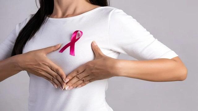
However, every woman can do the SADARI (breast self-examination) technique to detect lumps in her breasts.
SADARI should be done once a month, 7 to 10 days after the first day of menstruation.
The method is: Stand in front of a mirror and observe changes in the shape, size, skin color, and surface of the breasts.
Generally, the shape of the right and left breasts is not symmetrical, so there is no need to worry too much.
Raise both hands up and place them behind your head (nape).
Push your elbows back and forth while observing the shape and size of your breasts.
Feel the breast using three fingers (index, middle, and ring), then with gentle pressure.
Make a circular motion starting from the outside of the breast to the inside and touching the nipple.
Feel for lumps.
When bathing, place your right hand behind your head.
After soaping, check your right breast with your left hand in a circular motion, from the nipple to the outside of the breast.
Do the same on your left breast.
While lying down, place your left hand under your head.
Then, check your left breast with your right hand.
Do the same for your right breast.
Press both nipples and observe whether there is any abnormal fluid coming out from inside.
Another preventive measure is SADANIS (clinical breast examination), by medical personnel.
Every woman is advised to undergo SADANIS periodically, namely every 3 years for women aged 20 to 40 years and once a year for women over 40 years.
When to See a Doctor? It is advisable to have a check-up with a doctor if you find an abnormal lump that does not improve.
Especially if the breast looks bruised, the skin is reddish, and bleeding from the nipple.
.
Related Articles
health
3.60 கோடி லிட்டர் குடிநீரை சட்ட விரோதமாக திருடிய தனியார் கல்லூரிக்கு ரூ. 2 கோடி அபராதம் விதிக்க முடிவு!!
November 09, 2024

Home » 3.
60 கோடி லிட்டர் குடிநீரை சட்ட விரோதமாக திருடிய தனியார் கல்லூரிக்கு ரூ.
read morehealth
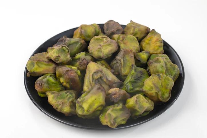
5 Health Benefits of Water Chestnuts
November 07, 2024

Water chestnuts, a popular ingredient in Asian cuisine, are not nuts but aquatic vegetables.
These crunchy, white orbs are the edible corms of a grass-like plant native to Southeast Asia, Southern China, Taiwan, Australia, Africa, and various islands in the Indian and Pacific oceans.
read more